The human knee is an engineering compromise. It needs to be stable enough to support your body weight (and then some — forces across the knee reach 2-3 times body weight during walking and 5-8 times during running or stair climbing) while also allowing a wide range of motion. The result is the largest, most complex joint in the body — and one of the most frequently injured.
About 25% of adults experience knee pain at any given time. It’s the second most common cause of chronic pain after low back pain. And the internet is full of conflicting advice: ice or heat, rest or exercise, surgery or supplements. Runner’s knee alone — a single condition — affects up to 25% of all runners and is the leading cause of visits to sports medicine clinics.
Here’s what most people get wrong about knee pain: they either catastrophize it (“my knee is destroyed, I need surgery”) or dismiss it (“it’s just wear and tear, nothing to do”). The reality is usually somewhere between those extremes, and the approach that works depends entirely on an accurate diagnosis.
Key Takeaways
- Patellofemoral pain syndrome (“runner’s knee”) is the most common cause of anterior knee pain and typically responds well to targeted physical therapy, not rest alone
- Meniscus tears are extremely common — found on MRI in up to 35% of asymptomatic people over 50 — so an MRI finding doesn’t necessarily explain your pain
- For most knee osteoarthritis, exercise is the single most effective treatment; it outperforms every supplement and many surgical procedures
- Knee surgery rates for many common conditions have decreased as evidence increasingly shows conservative treatment equals or beats surgical outcomes
- Pain with swelling after an acute injury (fall, twist, impact) warrants prompt medical evaluation — ligament tears, fractures, and locked knees need timely diagnosis
Anatomy Quick Reference
Understanding basic knee anatomy helps you make sense of diagnoses. The knee involves four bones (femur, tibia, fibula, patella), four major ligaments (ACL, PCL, MCL, LCL), two menisci (medial and lateral), multiple tendons, bursae, and the articular cartilage lining the joint surfaces.
Ligaments connect bone to bone and provide stability. The ACL (anterior cruciate ligament) prevents the tibia from sliding forward; the PCL prevents it from sliding backward. The MCL and LCL stabilize the inner and outer sides of the knee, respectively.
Menisci are C-shaped wedges of fibrocartilage that act as shock absorbers between the femur and tibia. They distribute load across the joint surface and improve stability.
Articular cartilage is the smooth, glassy coating on the ends of the femur, tibia, and underside of the patella. It allows near-frictionless movement. Once damaged, it has very limited ability to repair itself.
Tendons connect muscle to bone. The quadriceps tendon and patellar tendon (technically a ligament, since it connects two bones) are crucial for straightening the knee.
The Most Common Causes of Knee Pain
Patellofemoral Pain Syndrome (Runner’s Knee)
The most common cause of anterior (front) knee pain, particularly in runners, cyclists, and anyone who does a lot of knee bending. The pain is vague, aching, and located around or behind the kneecap. It worsens with stairs (especially going down), squatting, prolonged sitting (the “movie theater sign”), and running.
Despite its nickname, runner’s knee isn’t caused by running damaging the knee. It’s a tracking problem — the patella (kneecap) doesn’t glide smoothly in its groove on the femur, leading to irritation of the cartilage and surrounding tissues. Contributing factors include weak hip abductors (gluteus medius), tight IT band and quadriceps, overpronation of the foot, and sudden increases in training volume.
Treatment is exercise-based, not rest-based. The evidence overwhelmingly supports hip and quadriceps strengthening as the primary intervention. A 2018 Cochrane review confirmed that exercise therapy reduces pain and improves function, with hip-targeted exercises showing particular benefit. Specific exercises include clamshells, side-lying hip abduction, single-leg squats (partial range), terminal knee extensions, and step-ups.
What doesn’t work particularly well: knee braces (minimal evidence), complete rest (deconditioning worsens the problem), and cartilage supplements (glucosamine and chondroitin have consistently failed to demonstrate meaningful benefit for patellofemoral pain in high-quality trials).
Meniscus Tears
Meniscus tears are broadly divided into traumatic (acute injury, typically in younger patients) and degenerative (gradual wear, common in adults over 40). This distinction matters enormously for treatment decisions.
Traumatic meniscus tears — usually from twisting the knee under load — cause acute pain, swelling within hours, and sometimes mechanical symptoms: catching, locking (the knee gets stuck and won’t fully straighten), or giving way. Young patients with symptomatic traumatic tears, particularly those with mechanical locking, often benefit from arthroscopic surgery.
Degenerative meniscus tears are a different story. They’re incredibly common in middle-aged and older adults — MRI studies of asymptomatic volunteers find meniscal tears in 19% of people aged 50-59 and 56% of those aged 70-79. Having a meniscal tear on MRI does not mean the tear is causing your pain. This is one of the most important concepts in musculoskeletal medicine: imaging findings don’t always correlate with symptoms.
Multiple high-quality randomized trials — including the FIDELITY trial and the Finnish Degenerative Meniscal Lesion Study — have shown that arthroscopic partial meniscectomy (surgical trimming of a degenerative tear) provides no more benefit than sham surgery or physical therapy alone for degenerative tears without mechanical locking. Yet the surgery is still performed hundreds of thousands of times per year. The evidence has shifted practice, but slowly.
For degenerative meniscus tears, start with physical therapy, activity modification, and anti-inflammatory medications. Surgery should be reserved for tears causing true mechanical symptoms that don’t improve with 3-6 months of conservative treatment.
Osteoarthritis
Knee osteoarthritis (OA) affects roughly 14 million Americans, making it one of the most common joint diseases. It involves progressive loss of articular cartilage, along with changes to the underlying bone, joint capsule, and surrounding muscles. Contrary to the popular “wear and tear” explanation, OA is an active biological process involving inflammation, not just mechanical grinding.
Risk factors: age (prevalence increases sharply after 50), prior knee injury (ACL tear increases OA risk 3-5 fold), obesity (each pound of body weight adds roughly 4 pounds of force across the knee), female sex, genetics, and malalignment (knock-knees or bowlegs).
The hallmark symptoms: pain that’s worse with activity and better with rest (initially), stiffness after periods of inactivity (particularly morning stiffness lasting less than 30 minutes — longer suggests inflammatory arthritis), gradual loss of range of motion, and crepitus (grinding, cracking sounds).
Exercise is the single most effective non-surgical treatment for knee OA. This isn’t a nice-to-have recommendation — it’s first-line, evidence-level-A therapy. Strengthening the quadriceps and hip muscles reduces pain and improves function consistently across dozens of clinical trials. Low-impact aerobic exercise (cycling, swimming, walking) is equally important. The fear that exercise will “wear out” an arthritic knee is understandable but wrong — the knee does better with appropriate loading than with rest.
Weight loss is profoundly effective. The IDEA trial showed that a combination of exercise and diet-induced weight loss of at least 10% of body weight resulted in significant pain reduction and improved function in overweight adults with knee OA. Each pound of weight lost reduces the force across the knee by 4 pounds with each step.
Pharmacologically, topical NSAIDs (diclofenac gel) are recommended as first-line for mild-moderate OA with fewer systemic side effects than oral NSAIDs. Oral NSAIDs (ibuprofen, naproxen) are effective but carry GI and cardiovascular risks with long-term use. Acetaminophen was long recommended but recent evidence suggests it’s minimally effective for OA pain.
Corticosteroid injections provide short-term relief (weeks to a few months) but don’t modify the disease course. Frequent injections (more than 3-4 per year) may actually accelerate cartilage loss — a 2017 study in JAMA found greater cartilage loss with repeated steroid injections compared to placebo.
Hyaluronic acid injections (viscosupplementation) have a complicated evidence base. Meta-analyses reach conflicting conclusions depending on which trials they include. The American Academy of Orthopaedic Surgeons currently recommends against them, while some professional societies are more neutral. At best, the benefit is modest and comparable to placebo injection.
Total knee replacement is highly effective for severe, end-stage OA that hasn’t responded to conservative treatment. Patient satisfaction rates are around 80-85%. It’s a legitimate intervention when the time is right — but it’s major surgery with a serious recovery period, and it should not be the first thing you try.
IT Band Syndrome
The iliotibial band is a thick strip of fascia running from the hip to just below the outer knee. IT band syndrome causes sharp or burning pain on the outer (lateral) side of the knee, typically during running — characteristically at the same distance or time into a run. It’s the second most common running injury after runner’s knee.
Treatment focuses on addressing the root causes: hip weakness (again, gluteus medius is the usual suspect), training errors (too much too fast), and sometimes biomechanical issues. Foam rolling the IT band directly is widely practiced but may not be as helpful as commonly believed — the IT band is extremely tough connective tissue that’s nearly impossible to meaningfully stretch or elongate. Foam rolling the surrounding muscles (quads, TFL, glutes) makes more sense.
Ligament Injuries
ACL tears are one of the most well-known sports injuries. They typically occur from non-contact pivoting or landing — the classic basketball, soccer, or skiing mechanism. You feel or hear a pop, immediate swelling develops, and the knee feels unstable. ACL tears don’t heal on their own, but not everyone needs surgical reconstruction. Active individuals who want to return to cutting and pivoting sports generally benefit from reconstruction. Older adults or those who can modify their activities may function well with rehabilitation alone.
MCL sprains are the most common knee ligament injury. They usually heal without surgery because the MCL has a good blood supply. Treatment is bracing, rehabilitation, and time — 2-6 weeks for most sprains, longer for complete tears.
Bursitis
Prepatellar bursitis (“housemaid’s knee”) causes swelling directly over the kneecap — a visible, sometimes dramatic fluid pocket. It results from prolonged kneeling or direct impact. Most cases resolve with rest, ice, compression, and avoiding the aggravating activity. Occasionally, aspiration or antibiotics (if infected — red, warm, painful swelling with fever) are needed.
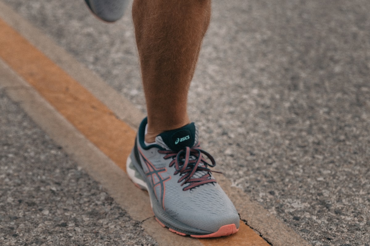
When Running Is and Isn’t the Problem
There’s a persistent myth that running destroys knees. The evidence says otherwise. A 2017 meta-analysis in the Journal of Orthopaedic & Sports Physical Medicine found that recreational runners actually had lower rates of knee and hip osteoarthritis (3.5%) than sedentary individuals (10.2%). Competitive/elite runners had higher rates (13.3%), suggesting a dose-response relationship, but the recreational running sweet spot appears to be protective, not destructive.
Running doesn’t damage healthy knees. Running on already-damaged knees, or running with significant biomechanical problems, or ramping up mileage too quickly — these cause problems. The “10% rule” (don’t increase weekly mileage by more than 10% per week) is a reasonable guideline, though not evidence-based as a specific number.
If you’re a runner dealing with knee pain, the most productive first step isn’t quitting running. It’s getting a proper assessment of hip strength, running biomechanics, and training load. Many running-related knee problems resolve with targeted strengthening without any change to running volume. For context on how this relates to other musculoskeletal issues, our article on lower back pain covers similar principles about the importance of movement over rest.
When to See a Doctor
Seek prompt evaluation if you experience:
- Significant swelling within hours of an injury (suggests ligament tear or fracture)
- Inability to bear weight on the affected leg
- Knee “locking” — you physically cannot straighten or bend it past a certain point
- Visible deformity
- Signs of infection: redness, warmth, fever combined with joint swelling
Schedule an appointment (not urgent but important) if:
- Pain persists beyond 2-3 weeks despite rest and self-care
- Pain interferes with daily activities or sleep
- You notice progressive instability or giving way
- Your knee pain is accompanied by systemic symptoms (fatigue, weight loss, morning stiffness lasting over 30 minutes in multiple joints — these suggest inflammatory arthritis). Diffuse bone aches combined with fatigue can also be among the signs of vitamin D deficiency, which a simple blood test can rule out
A note on imaging: you likely don’t need an MRI for most common knee problems. Clinical examination by an experienced provider is often sufficient to make a diagnosis. MRI is valuable for surgical planning, when the diagnosis is unclear, or when symptoms don’t respond to appropriate initial treatment. Getting an MRI “just to see” often reveals incidental findings (those degenerative meniscus tears) that create more anxiety than answers.
Frequently Asked Questions
Is cracking and popping in my knee dangerous?
Usually not. Crepitus (cracking, popping, grinding) without pain is extremely common and usually benign. It’s often caused by gas bubbles in synovial fluid or tendons snapping over bony prominences. Crepitus with pain is more significant and may indicate cartilage damage, but even then, the severity of the sound doesn’t correlate with the severity of the condition. Many people with advanced arthritis have quiet knees, and many people with perfectly healthy knees sound like a bowl of Rice Krispies.
Do glucosamine and chondroitin work for knee pain?
The GAIT trial — the largest and best-designed study — found that glucosamine and chondroitin, alone or in combination, were not significantly better than placebo for knee OA pain. Some subgroup analyses and smaller studies show modest benefits for moderate-to-severe OA, but the overall evidence is underwhelming. These supplements are generally safe, so there’s minimal harm in trying them for 3 months. But if you don’t notice improvement by then, stop wasting money.
Should I use ice or heat for knee pain?
Ice for acute injuries and inflammation (first 48-72 hours after an injury, or after activity that causes swelling). Heat for chronic stiffness and muscle tightness. For osteoarthritis, many patients find heat before activity (to loosen up) and ice after activity (to reduce inflammation) works well. This isn’t a settled science — the evidence for either intervention is surprisingly thin — so use what feels better. Neither will cause harm when used sensibly.
Can I still exercise with knee arthritis?
You should exercise with knee arthritis. This is not a contradiction — it’s a core evidence-based recommendation. Exercise reduces pain, improves function, and may slow disease progression. The key is choosing appropriate exercises (low-impact cardio, quadriceps and hip strengthening) and modifying intensity during flares. “Exercise hurts, so I stopped” is a common but counterproductive response. Initial discomfort during exercise is expected and generally doesn’t indicate damage. Pain that is significantly worse 24 hours after exercise suggests you did too much — scale back intensity rather than stopping entirely.
When is knee replacement surgery the right choice?
When knee OA is significantly limiting your quality of life despite adequate conservative treatment (exercise, weight management, medications, injections) over a sustained period. There’s no specific X-ray finding or age that determines the “right” time. The decision is driven by pain severity, functional limitation, and how much the problem affects your life. Most orthopedic surgeons recommend exhausting non-surgical options first, but also advise against waiting until you’re completely debilitated — outcomes are better when patients still have reasonable baseline function and fitness for recovery.